MIT.nano Seminar Series: January 2020

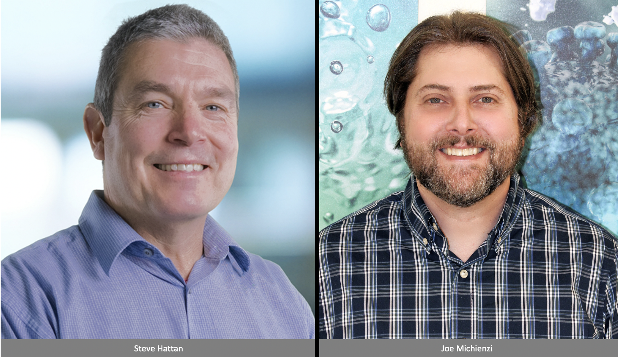
Steve Hattan, Ph.D., principal scientist, Waters Separations R&D Group
Joe Michienzi, principal research engineer, Waters Corporation
Date: Monday, January 27, 2020
Time: 3pm - 4pm; reception to follow
Location: Building 34, Room 401 (Grier Room)
This event is free and open to the public. Registration is required.
Abstract
Mass spectrometry has a role in changing the way we interrogate surfaces, providing molecular fingerprints with a level of information that enables artificial intelligence (AI) and machine learning (ML). In the biological space, this information is transforming diagnostic pathology by recognizing complex patterns in data and providing quantitative rather than qualitative assessments to oncologists.
Mass spectrometry has made significant advancements over the past decade in analytical speed, sensitivity, and robustness. These improvements have enabled in-roads for the technique in a broad array of scientific fields, including clinical oncology, where it has found application in the field of tissue imaging. Generally, mass spec imaging (MSI) refers to the systematic analysis of a surface resulting in a molecular map, or molecular image, of that surface. MSI has been employed in studies that monitor drug accumulation, clearance, and metabolism for numerous disease states to include cancer. The results of the technique compare favorably to current techniques such as radioluminescence microscopy.
To date, the most widely used technique for performing MSI (MALDI) requires a stringent sample preparation protocol prior to analysis, and the success and fidelity of this protocol is critical to the success of the procedure. Recent developments in novel ambient ionization methods, such as desorption electrospray ionization (DESI), have allowed for a more direct ionization and analysis of tissue surfaces. DESI enables investigation of samples free of chemical treatment and with minimal physical manipulation. The method uses a primary stream of charged droplets and high velocity gas (electrospray) to impinge a surface directly, dissolving analyte, and resulting in the ejection of secondary droplets that are collected and analyzed by the mass spectrometer. The DESI technique for surface analysis is rapidly evolving for in-situ analysis of samples ranging from tissues to non-biological materials such as plastics and metals providing detection and special resolution of critical compounds.
The achievable spatial resolution for DESI is largely determined by the dimensions of the divergent primary spray of solvent in relation to the tissue surface. Described here is a novel ESI source engineered to collimate and confine the electrospray stream with diameters of 20um, an order of magnitude improvement from the current technology. The confinement of the ion beam allows for high spatial resolution molecular images. Further advances will be required to achieve spatial resolutions in the 1um range and calls for a better understanding and simulations of ion and fluid manipulation and transport from a sample surface to the inlet of the mass spectrometer.
Recently, DESI combined with mass detection has been implemented for the direct analysis of thyroid cell biopsies with the aim of accurate detection and classification of cancerous/noncancerous cells and is a shining example of how MSI is impacting the world of medicine (PNAS October 22, 2019 116 (43) 21401-21408). The approach is to collect the output data from the analysis of cancerous/noncancerous cells, as determined by a pathologist, and build a molecular signature for each cell type. Having developed a reliable model, consensus spectra from classified cells can be compared against those gathered from routine biopsy. The results from these mass signature comparisons can not only boost the accuracy of the diagnosis, but also increase the speed and convenience of the analysis. The technology could have a large impact on the over 56,000 new patents who develop thyroid cancer each year (National Cancer Institute).
Our vision is to assemble an integrated system that would enable this technology to become an industry standard in pathology. This presentation will share an overview of the status of several elements that make up this system as well as discuss some of the challenges still faced in bringing it to a level of functionality for the research and long-term clinical market.
Biographies
Stephen Hattan, Ph.D., is principal scientist in Waters Separations R&D group. He received his Ph.D. in analytical chemistry in 1996 from the University of New Hampshire. His thesis work focused on protein purification and characterization. He completed a postdoc at the Wadsworth Center, NY State Dept. of Health in Albany, NY, characterizing glycan structures by NMR and a second postdoc at the Whitehead Institute MIT Cambridge, MA, in the laboratory of Dr. Paul Matsudaira doing protein sequencing by mass spectrometry prior to joining Genetics Institute 1998. After the completion of the human genome, Dr. Hattan was hired as a lead separations scientist at Celera in its newly formed proteomics department. He has since spent ~20 years working at the interface between separation science and mass spectrometry at Applied Biosystems, SimulTOF systems and now Waters Corporation where he is assigned to the global mass spec imaging research team. His efforts at Waters have been aimed at hardware improvements to the desorption electrospray ionization source (DESI source).
Joe Michienzi has been, for the past twenty years, helping biologists, chemists, and engineers succeed through innovation of mechanical and electrical systems. Joe started his career working as a research engineer on a NIH-funded program to reduce DNA sequencing costs for the Human Genome Project. Joe graduated from Wentworth Institute of Technology with a mechanical engineering degree. He joined Waters Corporation in 2003 while earning his BS in electrical engineering from Northeastern University. Joe has been part of many important technological breakthroughs during his time at Waters, most notably for his development of a novel ceramic microfluidic device for the ionKey/MS™ system which won a top innovation award from The Analytical Scientist in 2014 and an R&D 100 Award in 2015. Joe has earned numerous patents and authored publications over his career in the areas of microfluidics and liquid, gas, and supercritical fluid chromatography. Joe is passionate about research in science and engineering. He currently manages a team of research engineers focused on developing innovative technologies to solve intractable problems in separation science.
